To minimize excess ssDNA in the final structure, which may result in nonspecific object aggregation or otherwise interfere with folding, we decided to perform asymmetric PCR ( αPCR ) to generate object-specific scaffolds for folding.
αPCR was performed with a sense primer concentration of 1 mM, an anti-sense primer concentration of 20 nM, 50 ng of M13mp18 dsDNA template, 200 mM dNTPs mixed in a final volume of 50 μl and 1 unit of Q5TM High-Fidelity DNA Polymerase(NEB). The aPCR program used is as follows: 95°C, 4 min for the initial denaturation; followed by 35 to 40 cycles of 95°C, 30 s; 55° , 30 s; 72°C, 5min; 72℃for final extention, and hold at 4℃.
Identification and purification
The aPCR products were run through 0.8% low-melting temperature agarose gel ( 0.2g agarose, 25ml 1× TAE), under a constant voltage of 80V. The products were extracted and purified by Gel DNA recovery kit.
Origami reaction protocol
Prepare the reaction
All staples were diluted with ddH2O to a concentration of 500 nM. Scaffold is mixed with 10 times concentration of staple strands in 1× TAE-Mg2+. You can see the details in Table 1
Table 1
Concentration
[μl]
Final concentration
Scaffold
10~20 nM
25
5~10 nM
Staple
167~333 nM
15
50~100 nM
10X TAE
——
5
1× TAE
10× Mg2+ buffer
100~200 mM
5
10~20 mM
Annealing
The temperature was lowered from 80 ℃ to 60℃ at 4min ℃-1, then from 60℃ to 24℃ at 20 min/℃-1 .
Hold at 4℃
Structural change
Expansion
Take 25μl Origami sample and mix with 25μl fuel strand solution, which is 15 times the concentration of the Origami products.
Incubate at 37℃ for 30 min, then cool to 15℃ by 3℃/min.
Constriction
Take 25μl expansion sample and mix with 25μl anti-fuel strand solution, making the fuel strand: anti-fuel strand ratio of 5:1.
Incubate at 37℃ for 30 min, then cool to 15℃ by 3℃/min.
Purification of Origami products
The aPCR products were run through 2% low-melting temperature agarose gel (0.5 g agarose, 25ml 1× TAE) under a constant voltage of 80V, then be extracted and purified with Gel DNA recovery kit(厂名).
AFM protocol
Take a 5μl volume of the sample for AFM, let it be deposited onto a freshly cleaved mica and left to adsorb to the surface for 5 min. Then wash with ddH2O to remove the salt, allow it to air dry and wait for imaging.
The DNA samples were imaged in tapping mode.
TEM protocol
Drop 5μl of the sample solution on the grid and left to adsorb for 5 min. For staining, the grid was touched with a drop of 2% uranyl acetate solution for 1min. Let the grid dry and keep it at room temperature.
Dynamic Light Scattering
For the measurement, 100μl sample solutions were measured at a concentration of 10 nM in 1× TAE Buffer. The sample temperature was maintained at 25℃ during measurement.
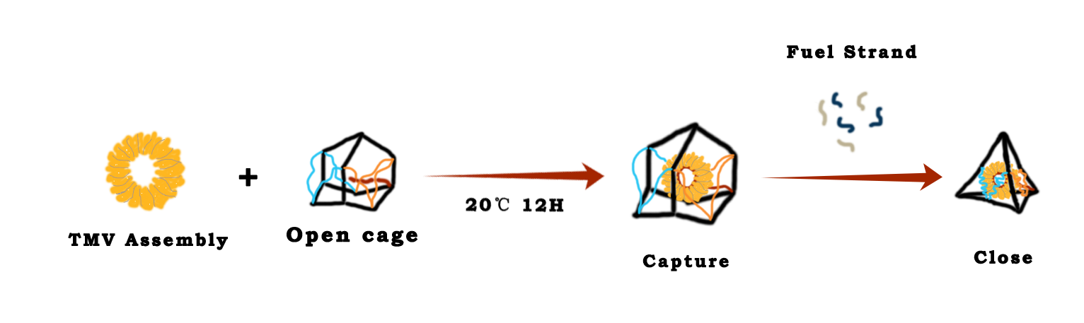
Assembly of TMV coat protein(CP)
The TMV coat proteins purified by DEAE anion exchange chromatography were disaggregated through dialyzing in the PBS buffer (PH 8.0) at 4℃ for 48h, and for assembly of TMV disc, the disaggregated proteins were dialyzed in PBS buffer (PH 7.0) at 4℃ for more than 24h.
Capture of the TMV disc
The assembled TMV disc were incubated with the open cage at 20℃,12h. Then the mixture was added with anti-fuel strands and incubating for another 40 min to close the cage.